Notes from the receiving surgeon
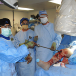
My initial plan involved retrograde intramedullary nailing (IMN) the right femur immediately. I debated external fixation versus a distal femoral traction pin for the left femur. If Khan remained stable, I planned on antegrade IMN of the left femur in 24 to 48 hours and open reduction, internal fixation (ORIF) of the left ankle and metatarsals in 7 to 10 days.
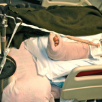
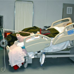
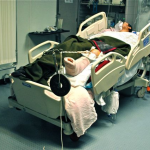
Safely “stabilizing” long bone fractures in the field can be challenging without traditional equipment. I did want to treat the left femur fracture with an intramedullary device sooner rather than later and did not want to have external fixation pins in the proximal segment if I did not need to wait. I therefore placed a distal femoral traction pin and improvised skeletal traction with some borrowed weights. When doing so, because we did not have standard frames, slings, and pulleys for balanced traction, I used padding to support the thigh and leg and elevated the vector of traction to try to keep it “in line” with the femur.
Postoperative day 2
Overnight nursing reported Khan being “oxygen dependent.” He desaturates very quickly when weaned from oxygen. A work up showed:
- Khan was a 2 pack per day smoker
- Arterial blood gas (ABG) on 50% FiO2 showed hypoxemia with mild shunting
- A sonosite showed femoral veins are “compressible”
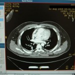
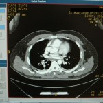
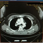
I wondered if this indicated a differential diagnosis of pulmonary embolism (PE), fat embolism syndrome (FES), chronic obstructive pulmonary disease (COPD), or a combination of all three. Fortunately, a contrast CT was available and I diagnosed a PE.
We heparinized him immediately, but Khan could not be transported to a higher level hospital. We were to be his definitive care facility. Fortunately, he remained hemodynamically stable despite the pulmonary embolism. Unfortunately, we did not have an interventional radiologist nor a vascular surgeon assigned to our facility. I considered options of definitive fixation with external fixation alone, which could be done through a short window in his heparin therapy with percutaneous technique by sending large external fixation pins up the femoral neck in conjunction with shaft pins to maximize the fixation in the proximal segment. Trying to ream for immediate IMN in this setting would likely be too risky, as the emboli to his lungs might push him over the edge of cardiopulmonary stability. Any additional surgeries during full medical anticoagulation might allow for large hematoma formation.
What would be your next steps as the treating surgeon? Check back to learn how the surgeon proceeded in this austere environment.


