Self-Inflicted Gunshot Wound to the Hand in Partner Forces – Part 2

Share

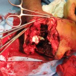
Intra operative

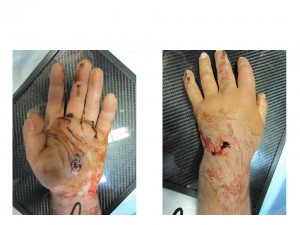
  • Incise and drain entrance and exit wounds
  • Open carpal tunnel release
  • Intact median nerve
  • Intact extensor tendons, < 50% damage to flexor digitorum superficialis and flexor digitorum profundus
  • Intact deep and superficial palmar arch
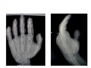
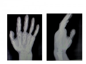
  • Comminuted third metacarpal and capitate fracture, non-displaced fourth metacarpal base fracture
  • Surgical incisions closed loosely, entrance/exit wounds left open

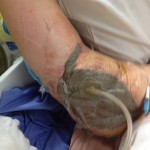
 Post operative

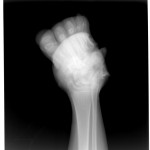
fig 3

If you could not guarantee continuity of care (must pass care to a lower standard), what instructions would you give to the accepting physician and the patient?

Are there any cultural/military discipline implications for this injury?

Self-Inflicted Gun Shot Wound to the Hand in Partner Forces – Part 1

Share

Background

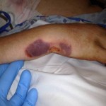
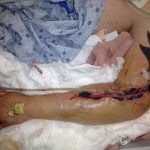
  • 26-year-old right hand dominant male Afghan, member of the Afghan Local Police (ALP).
  • Self-inflicted gun shot wound to left hand, presented for treatment within 8 hours to the Forward Operating Base surgical team.
  • Decreased sensation median nerve distribution, intact radial and ulnar nerve sensation.
  • Brisk capillary refill was less than 2 seconds inall digits (perfusion adequate). 
  • Motor exam not possible due to pain.

Pre operative

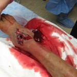
fig 1

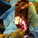
fig 2

Operation proceeded within an hour after presentation. 

In the austere environment, what should be done for this patient?

What are the goals of surgery?

Orthopaedics in Motion – Volume 3, Number 3 – Fall 2013

Share

Now available online.

In This Issue:

  • When Plane-Crash Victims Arrived at Stanford Medicine, Response Teams Were Ready
  • Traveling Abroad This Fall? Be Prepared Before You Go.
  • Refresh Your Trauma Skills (part four of a series)
  • Disaster Response Course
  • Don’t Overlook These Basics: 5 Ways Your Culture Can Improve Patient Satisfaction
  • Editorial: The Lowest Common Denominator and a Firm Grasp of the Obvious
Orthopaedics in Motion is a quarterly newsletter containing sections on disaster preparedness and trauma care. The newsletter also includes overviews of the latest aspects of clinic care, continuing education, research and innovation pertinent to the practicing orthopaedic surgeon.

Pediatric Blast Injury: Bilateral Amputation – Part 2

Share

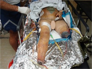
The surgical team performed external fixation of the right femur, followed by debridement and irrigation of Masooma K.’s lower extremities, followed by a completion amputation to the viable level of her left lower extremity, above the knee.

“I debated limb salvage versus completion amputation to the patient’s right lower extremity.  While my partners were working on her abdomen and I was completing her left leg amputation, the patient began to have difficulty ventilating and displayed tension physiology, so we placed a second chest tube.”

“We began to have trouble keeping the patient’s blood pressure stable.  Her abdomen became edematous after her intestinal repairs, so I elected to amputate her right lower extremity at a very distal ankle disarticulation.”  Masooma K. survived the night and stabilized later postop day 1, enough for aeromedical critical care transportation to a higher echelon facility.

fig 6

1. Was there any real role for limb salvage in an austere environment, given that this patient would be transferred to a higher level hospital within 24 hours?

2. Is there any difference between a Syme amputation vs. a low transtibial amputation vs. a below-knee amputation in countries supported solely by the International Committee of the Red Cross for prosthetics?

3. What is the fate of a female double amputee in Afghanistan?

Pediatric Blast Injury: Bilateral Amputations – Part 1

Share

Masooma K. and her family were returning home and became caught between American and Taliban fighters just as an explosion went off.

“A 13-year-old female was Medevac’d to our forward surgical team.  She had been injured in the blast and suffered penetrating abdominal, chest, and bilateral lower extremity wounds,” a surgeon observed. The forward surgical team consisted of a general surgeon, orthopaedic surgeon, CRNA, OR nurse, and OR tech with limited radiologic (single shot portable X-ray only) and lab support.  Blood products were available, but overnight holding capability only was a severe limitation.  Trauma stabilization surgery was limited given the austere environment and limited staff/equipment.

fig 1

Masooma K. had an initial blood pressure of 80/43 and a pulse of 126.  She had decreased left chest breath sounds and an oxygen saturation of 88% with a small penetrating wound on the left flank.  Her left leg was mangled and unsalvageable due to lack of blood flow distal to her intraarticular knee fracture, when assessed with the tourniquet down, and a degloving injury of tissue off the popliteal fossa.  Masooma K.’s mangled right foot had massive soft tissue loss and a non-viable heel pad.  Clinical evaluation showed a fracture dislocation of the subtalar joint, with > 50% loss of the calcaneus with extrusion and loss of the talar head fracture.  Additionally, there was gross instability of the right thigh.  Intraoperative radiographs showed a middle third femoral shaft fracture.

fig 2 fig 3 fig 4 fig 5

“We intubated her in the trauma bay and placed a left chest tube.  Then we took her emergently to the operating room for a damage control exploratory laparotomy, due to her penetrating abdominal wound.”

What are your orthopaedic priorities at this point?  In the austere environment given your team’s resources and the possibility that you may take more trauma, what would you elect to do surgically for this adolescent girl?

Orthopaedics in Motion – Volume 3, Number 2 – Summer 2013

Share

Orthopaedics in Motion – Volume 3, Number 2 – Summer 2013 is now available online.

Orthopaedics in Motion is a quarterly newsletter containing sections on disaster preparedness and trauma care. The newsletter also includes overviews of the latest aspects of clinic care, continuing education, research and innovation pertinent to the practicing orthopaedic surgeon.

In This Issue:

Orthopedic Surgeon Dr. John Cowin Rushed to Help Victims of Boston Marathon Bombs

Recruitment Resolutions: Avoid Common Hiring Pitfalls

Refresh Your Trauma Skills (part three of a series)

Healthy Summer Foods

Editorial: Preparing for Disaster

Orthopaedics in Motion – Volume 3, Number 1 – Spring 2013

Share

Orthopaedics in Motion – Volume 3, Number 1 – Spring 2013 is now available online.

Orthopaedics in Motion is a quarterly newsletter containing sections on disaster preparedness and trauma care. The newsletter also includes overviews of the latest aspects of clinic care, continuing education, research and innovation pertinent to the practicing orthopaedic surgeon.

Orthopaedics in Motion – Volume 2, Number 4 – Winter 2012

Share

Orthopaedics in Motion – Volume 2, Number 4 – Winter 2012 is now available online.

Orthopaedics in Motion is a quarterly newsletter containing sections on disaster preparedness and trauma care. The newsletter also includes overviews of the latest aspects of clinic care, continuing education, research and innovation pertinent to the practicing orthopaedic surgeon.

Multiple Gunshot Wounds – Part 2

Share

On the day of the injury, the trauma surgical team immediately initiated advanced trauma life support stabilization protocols for trauma resuscitation. They achieved intravenous access and initiated shock treatment. A warmed operating room had been prepared and blood products were ordered for planned rapid infusion. The team placed a chest tube and confirmed the diagnosis of a simple pneumothorax from a penetrating gunshot wound. They performed a brief neurovascular exam of both hands, which suggested Smith suffered no major arterial disruption and all major nerve distributions were sensate. While prepping Smith in the operating theater, the team performed rapid sequence intubation for anesthetic care. The general surgeons performed an exploratory laparotomy while the orthopaedic surgeons performed irrigation and debridement of both upper extremity injuries and multiple soft tissue wounds, and applied negative pressure wound dressing. The surgeons took care to ensure that any penetration to the elbow joint was ruled out via saline arthrogram and direct inspection.

On the second day, the orthopaedic surgeons repeated irrigation and debridement of Smith’s wounds.

Within 72 hours of his injuries, Smith was stabilized and aeromedically evacuated from the theater with both of his arms splinted. He had been extubated and was able to follow commands and demonstrated use of both hands.

He arrived at a hospital outside the combat zone and was recovering well from his chest and abdominal wounds. Surgeons continued repeat irrigation and debridement of his open wounds. Five days after he was injured, Smith arrived at a stateside tertiary care military hospital and received repeated irrigation and debridement of his wounds.

At this point, what plan would you recommend for definitive care of Smith’s injuries? What concerns would you have for the positioning and approach?  Would you fix both sides at once?

Multiple Gunshot Wounds – Part 1 Now Live

Share

Maj Smith, a 22-year-old male, was just three weeks into his second tour of duty. While on foot patrol one afternoon, Smith and his squad broke off from the company to begin house-to-house searches in one of the more dangerous neighborhoods in the area. Moments after entering one home, they surprised a group of insurgents, who quickly open fire on the squad.

“I remember ordering my men to take cover and the next thing I knew, I was in a battalion aid station,” recalled Smith.

Smith suffered multiple gunshot wounds while trying to get his squad to safety. A nearby squad heard the gunfire and was soon able to contain the insurgents and transport Smith and his men to medical care. Both of Smith’s arms were wounded and he was bleeding from his flank, where a bullet penetrated both his right arm and his chest. Also, he was having difficulty breathing. The company had been well trained in front-line first aid and buddy care. One soldier applied tourniquets to both upper arms to slow the bleeding while another applied direct pressure and clean bandages to Smith’s open wounds. They also performed decompression of Smith’s right chest, which eased his breathing while they waited for the medevac chopper to arrive.

At a nearby combat support hospital, Smith saw the first doctor to manage his many wounds. The diagnosis included orthopaedic injuries consisting of bilateral distal third, open, comminuted humerus fractures, a left small finger comminuted metacarpal fracture, and multiple soft tissue fragmentary injuries. Non-orthopaedic injuries were a right-side pneumothorax, bilateral testicular rupture, and a gunshot wound to the abdomen.

In an echelon 2 medical facility, where basic surgical stabilization care can be provided, what sequence of care would you recommend for this patient?