Orthopaedics in Motion – Volume 3, Number 3 – Fall 2013

Share

Now available online.

In This Issue:

  • When Plane-Crash Victims Arrived at Stanford Medicine, Response Teams Were Ready
  • Traveling Abroad This Fall? Be Prepared Before You Go.
  • Refresh Your Trauma Skills (part four of a series)
  • Disaster Response Course
  • Don’t Overlook These Basics: 5 Ways Your Culture Can Improve Patient Satisfaction
  • Editorial: The Lowest Common Denominator and a Firm Grasp of the Obvious
Orthopaedics in Motion is a quarterly newsletter containing sections on disaster preparedness and trauma care. The newsletter also includes overviews of the latest aspects of clinic care, continuing education, research and innovation pertinent to the practicing orthopaedic surgeon.

Orthopaedics in Motion – Volume 3, Number 2 – Summer 2013

Share

Orthopaedics in Motion – Volume 3, Number 2 – Summer 2013 is now available online.

Orthopaedics in Motion is a quarterly newsletter containing sections on disaster preparedness and trauma care. The newsletter also includes overviews of the latest aspects of clinic care, continuing education, research and innovation pertinent to the practicing orthopaedic surgeon.

In This Issue:

Orthopedic Surgeon Dr. John Cowin Rushed to Help Victims of Boston Marathon Bombs

Recruitment Resolutions: Avoid Common Hiring Pitfalls

Refresh Your Trauma Skills (part three of a series)

Healthy Summer Foods

Editorial: Preparing for Disaster

Orthopaedics in Motion – Volume 3, Number 1 – Spring 2013

Share

Orthopaedics in Motion – Volume 3, Number 1 – Spring 2013 is now available online.

Orthopaedics in Motion is a quarterly newsletter containing sections on disaster preparedness and trauma care. The newsletter also includes overviews of the latest aspects of clinic care, continuing education, research and innovation pertinent to the practicing orthopaedic surgeon.

Orthopaedics in Motion – Volume 2, Number 4 – Winter 2012

Share

Orthopaedics in Motion – Volume 2, Number 4 – Winter 2012 is now available online.

Orthopaedics in Motion is a quarterly newsletter containing sections on disaster preparedness and trauma care. The newsletter also includes overviews of the latest aspects of clinic care, continuing education, research and innovation pertinent to the practicing orthopaedic surgeon.

Orthopaedics in Motion – Volume 2, Number 3 – Fall 2012

Share

Orthopaedics in Motion – Volume 2, Number 3 – Fall 2012 is now available online.

Orthopaedics in Motion is a quarterly newsletter containing sections on disaster preparedness and trauma care. The newsletter also includes overviews of the latest aspects of clinic care, continuing education, research and innovation pertinent to the practicing orthopaedic surgeon.

Orthopaedics in Motion – Volume 2, Number 2 – Summer 2012

Share

Orthopaedics in Motion – Volume 2, Number 2 – Summer 2012 is now available online.

Orthopaedics in Motion is a quarterly newsletter containing sections on disaster preparedness and trauma care. The newsletter also includes overviews of the latest aspects of clinic care, continuing education, research and innovation pertinent to the practicing orthopaedic surgeon.

Case of the Month — Spanning Fixation and Proximal Femur

Share

M. Aziz, a 52-year-old male Afghan civilian, sustained a right tibia shaft fracture, a left tibial plateau fracture, and a left subtrochanteric fracture after driving over an improvised explosive device while on his way to visit his brother.
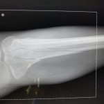
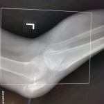
      

Question:  How would you address these closed injuries in an austere environment with only external fixation available for definitive treatment?

“I used external fixation for the right tibial shaft fracture.  It was an easy, straightforward procedure,” explained Aziz’s surgeon.

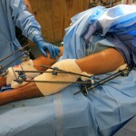
The damage to the left side was more complicated. “I was running low on external fixation connectors, pins, and rods with the case, so I decided to use the distal femur pins in both the left spanning knee fixation and the left hip fixation,” said the surgeon.

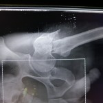
As the operating room does not have a fluoroscope, prior to surgery the surgeon places a radio-opaque marker next to a mark he makes on the patient’s skin or next to an entry wound, such as a bullet hole.  He then uses images from his own camera to determine where to place the pins based on the relationship of the marker to the patient’s anatomic landmarks.

“One trick I like is to place a flat plate under the hip before the patient is prepped.  That way, after inserting the first pin blind with no C-arm, I can take an X-ray and check a plain film to see what adjustments need to be made and where the second pin should be placed in relation to the first.  This saves a little bit of time, as we do not have to wait for a plate to be placed.”

In Aziz’s case, the surgeon did not want to place two additional pins in the patient’s femur.  He was running low on external fixator pins and wanted to reduce the number of pins placed, since the patient already had multiple pins placed.  The surgeon used the same pins in the distal femur for the hip and proximally for the spanning knee fixation.

First, he stabilized the knee so that construct could be used to reduce the hip once the proximal pins were placed. For reduction of the knee, the surgeon pulled in extension, allowing for slight flexion of the knee. He “cheated” valgus a little past what he believed was aligned, as he did not have a C-arm in the operating room and had to use plain films to verify each reduction attempt (these injuries have a tendency to fall into varus), and was successful.

For the hip, the surgeon pulled longitudinal traction distally and “did the opposite of what the proximal fragment wants to do.” He extended, adducted, and internally rotated the proximal fragment to achieve reduction and again, it worked.  For a final check, he consulted the X-ray.
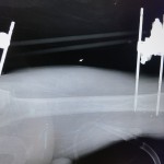
      

“When I do the reductions I tighten one set of connectors and then perform the reduction with my assistant ready to tighten the second set. For the knee I tightened the proximal first and then reduced and tightened the distal. For the hip it was the opposite,” the surgeon explained.

Aziz required no blood products during surgery. Within one hour of the surgery being completed, the dust-off crew medevaced him to a civilian hospital.  He was transferred in stable condition.

Case of the Month — Above-Knee Amputation Now Live

Share

Ara K., a 14-year-old girl, along with her sister and cousin, were riding in a civilian bus when it ran over an improvised explosive device (IED). Although the site had recently been discovered and cleared, a new IED had already been replaced in the same location.

All passengers on Ara K.’s side of the bus were injured or killed. Doctors attempted to revive her cousin at the forward operating base, but both he and Ara K.’s sister died.

Ara K. suffered massive damage to both legs. Surgeons performed a below-knee amputation on her right leg, but her left leg was more severely damaged. With skin and soft tissue loss, a below-knee amputation was not feasible for her left leg. Posteriorly, large areas would have had near-exposed bone because there was not enough tissue for coverage.

During hurried discussion, one of her surgeons stated, “If we shunted the left leg, it would have been to a high below-knee amputation [due to the soft tissue damage around the knee] and I don’t know how helpful that would be.”

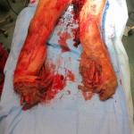
Surgeons ultimately decided to perform an above-knee amputation. Ara K.’s injuries were so proximal, surgeons obtained control of her femoral artery just proximal to the bifurcation and passed vessels loops around it in case she started to hemorrhage massively from her wounds.  Care was taken to keep the tourniquet in place and prep it into the surgical field so that controlled release during surgery could be performed.

Ara K. had damaged and thrombosed vasculature just distal to the level where surgeons made the bony amputation cut. The surgeons had to take more muscle medially in order to debride her injury appropriately. They decided to retain as much viable muscle as possible together with her flap, but in the end had to shorten the femoral cut even more because of concerns that the flap was too thin over the end of the femur.  They felt that leaving only viable skin over the distal stump would lead to more complications with eventual prosthesis fitting.

In the operating room, Ara K. was given 1:1 transfusions of fresh frozen plasma and packed red blood cells, and surgeons did not have to use proximal control to stop the bleeding.

Ara K. was maintained for a week at the forward base hospital for stabilization of both her lower extremity injuries and other issues related to the blast.  She was ultimately transferred in stable condition to the local hospital for completion of care.

In above knee amputations, what is the utility of myodesis with consideration of future mechanical function and prosthesis fitting?

In rural Afghanistan, like most austere environments, prosthesis fitting is quite difficult.  There is a national program that is provided, but the waiting list can be quite long.  If NO prosthesis services are available, would that change surgeons’ preferences for amputation level decision making?  In this case would you have given the “high below-knee amputation” a chance before converting to an above-knee amputation?

Orthopaedics in Motion – Volume 2, Number 1 – Spring 2012 is now available online.

Share

Orthopaedics in Motion – Volume 2, Number 1 – Spring 2012 is now available online.

Orthopaedics in Motion is a quarterly newsletter containing sections on disaster preparedness and trauma care. The newsletter also includes overviews of the latest aspects of clinic care, continuing education, research and innovation pertinent to the practicing orthopaedic surgeon.

For Orthopaedics in Motion – Volume 1, Number 4 – Winter 2011 please visit The Disaster Preparedness Toolbox and click the Newsletter link.

For Orthopaedics in Motion – Volume 1, Number 3 – Fall 2011 please visit The Disaster Preparedness Toolbox and click the Newsletter link.

For Orthopaedics in Motion – Volume 1, Number 2 – Summer 2011 please visit The Disaster Preparedness Toolbox and click the Newsletter link.

For Orthopaedics in Motion – Volume 1, Number 1 – Spring 2011 please visit The Disaster Preparedness Toolbox and click the Newsletter link.

Case of the Month – Hip Injury – Part 2 Now Live

Share

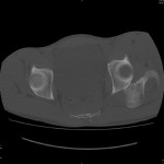
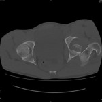
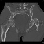
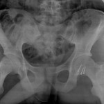
The surgeon immediately recognized that Spc Walsh had a closed left hip fracture dislocation and without delay attempted a manual closed reduction. Unfortunately, this was not successful, even with the patient under anesthesia. The surgeon then attempted another closed reduction, this time with a Steinmann pin placed in the proximal femur. The fracture, however, was found to be irreducible.

The surgeon decided to perform an open reduction with internal fixation of the Pipkin 2 femoral head fracture.

figure 5

The patient was then transported by aeromedical evacuation back to his home country where he reportedly went on to complete his healing and recovery without significant complication.